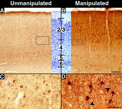
Figure 3.
Effects of subplate ablation on GAD expression. (A–D) GAD-67 immunostaining in primary visual cortex at P28 after injection of kainic acid into the subplate on P7. (A) GAD-67 immunostaining in the hemisphere contralateral to the kainic acid injection, showing levels of staining comparable to that of unmanipulated control animals. (B) GAD-67 immunostaining in visual cortex overlying injection sites in the subplate-ablated hemisphere from the same animal shown in A. Neuropil immunostaining is significantly increased throughout the cortical plate and in the underlying white matter. (C and D) High-magnification photomicrographs of the boxed areas at the border of layers 3 and 4 in A and B. Immunopositive cells are larger and more intensely labeled in the manipulated D than the unmanipulated C hemisphere. The middle portions of A and B represent cresyl violet counterstained sections adjacent to stained sections. Heavily labeled processes are visible (arrowheads) in D. Numbers denote cortical layers. Bars = (A and B), 200 μm; (C and D), 50 μm.

