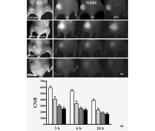
Figure 3.
FRI of different tumour xenografts over time. (a) White light images of different tumour xenografts (as indicated), followed by fluorescence reflectance imaging (FRI) images taken sequentially after application of 2 μmol/kg body weight SIDAG. Note the strong fluorescence signal in the HT1080 (n = 11) fibrosarcomas and in the MDA-MB435 (n = 10) melanomas, whereas the MCF7 (n = 9) and DU4475 (n = 13) adenocarcinomas exhibit only moderate tumour fluorescence. (b) Quantitative data analysis revealed significantly higher contrast to noise ratios (CNRs) for the HT180 and MDA-MB435 xenografts compared with the MCF7 and the DU4475 tumours.

