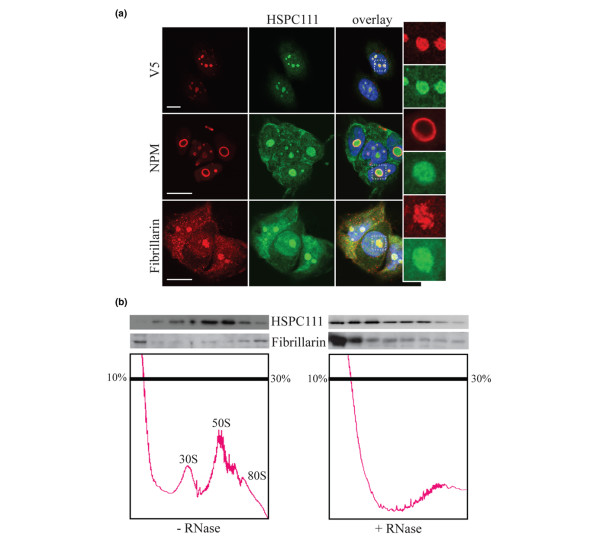
Figure 3.
HSPC111 resides in high molecular weight protein complexes in the nucleolus. (a) Detection of endogenous and tagged HSPC111 by indirect immunofluorescence. Upper panels: Immunostaining of MCF-7/HSPC-NV5 cells with purified antibodies against endogenous protein (HSPC111; green) and the V5 tag (V5; red). Middle panels: Parental MCF-7 cells were stained with anti-HSPC111 (green) and anti-nucleophosmin (NPM; red) antibodies. Lower panels: Parental MCF-7 cells stained with anti-HSPC111 (green) and anti-fibrillarin (red) antibodies. DNA was counterstained with DAPI (4,6-diamidino-2-phenylindole; blue). Images are representative of at least two independent experiments. Bar = 10 μm. (b) Nuclear extracts of MCF-7 cells treated with or without RNase A were fractionated on sucrose density gradients. The trace from continuous monitoring of absorbance at 254 nm is shown. Fractions were precipitated and immunoblotted for HSPC111 and fibrillarin.

