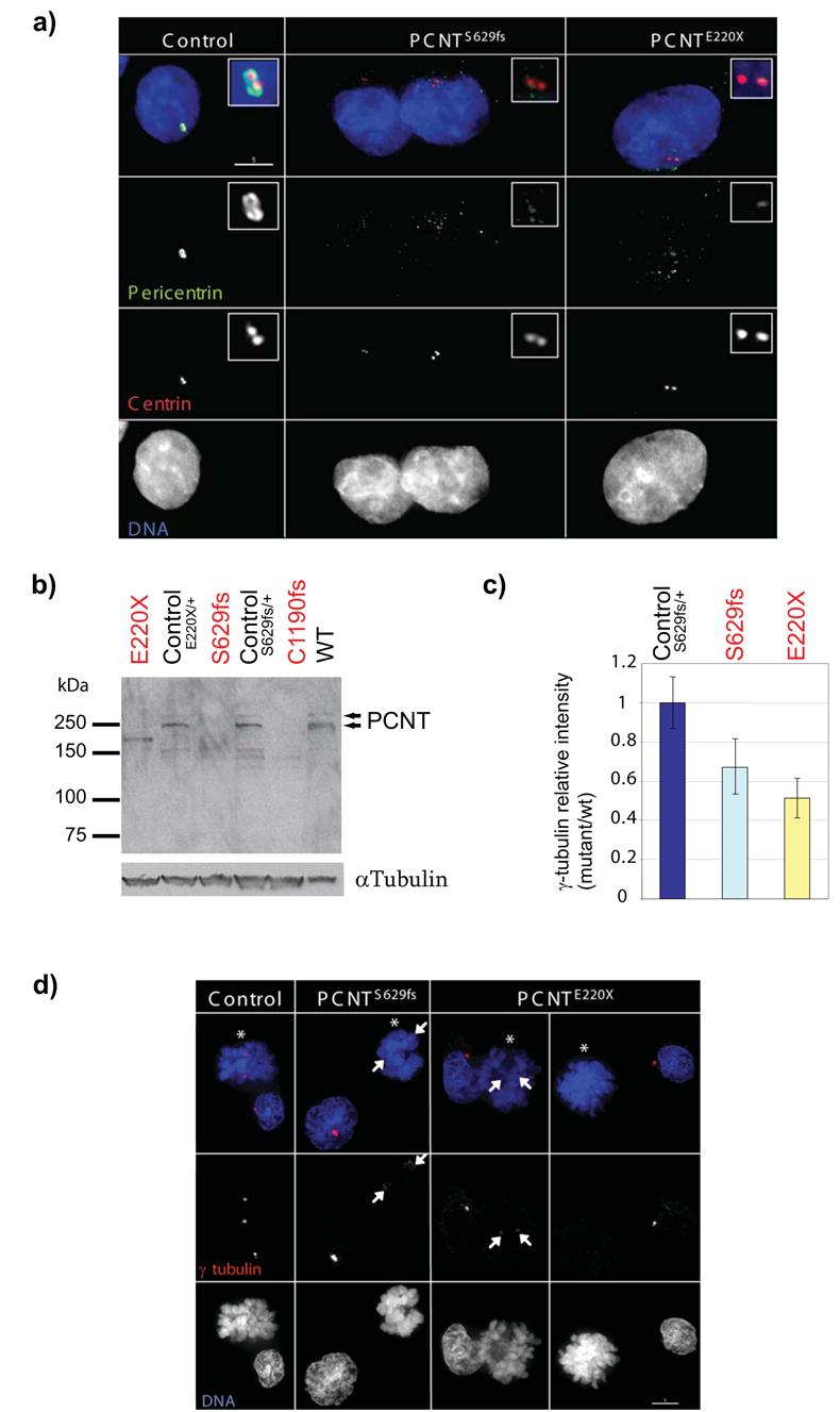
Figure 2. Pericentrin localisation and function is disrupted in PCNT-Seckel cell lines.

(a) Pericentrin is not localised to the pericentiolar material in PCNT-Seckel cells. Deconvolved immunofluorescent images from PCNT-Seckel and control lymphoblastoid cell lines. Pericentrin (ab448-100, green) and centrin (red). Scale bar 5μm. Control, heterozygote relative, PCNT220X/+. (b) Immunoblot of LCL cell lysates with Pericentrin ab448-100 antibody, that detects both Pericentrin A and B isoforms. Two pericentrin (arrowheads) isoforms are absent from PCNT-Seckel cells, but present in control lymphoblastoid cells from heterozygous relatives. A smaller protein product of ∼170kD is detected in the PCNT220X cell line,that might represent an aberrant truncated PCNT protein product. Loading control, alpha-tubulin. (c,d) γ–tubulin localisation is frequently reduced or absent during mitosis in PCNT-Seckel cells. (c) Quantification of γ-tubulin signal in PCNT-Seckel mitotic cells at prometaphase and metaphase, relative to wild-type. n=20. error bar, s.d. γ-tubulin signal intensity is significantly reduced relative to wild type cells (p<0.05, S629fs; p<0.001, E220X) (d) In PCNT-Seckel mitotic cells (astericks), γ–tubulin centrosomal staining is reduced (arrows) or absent, relative to centrosomal staining in adjacent interphase cells, or in control cells from a heterozygous relative.
