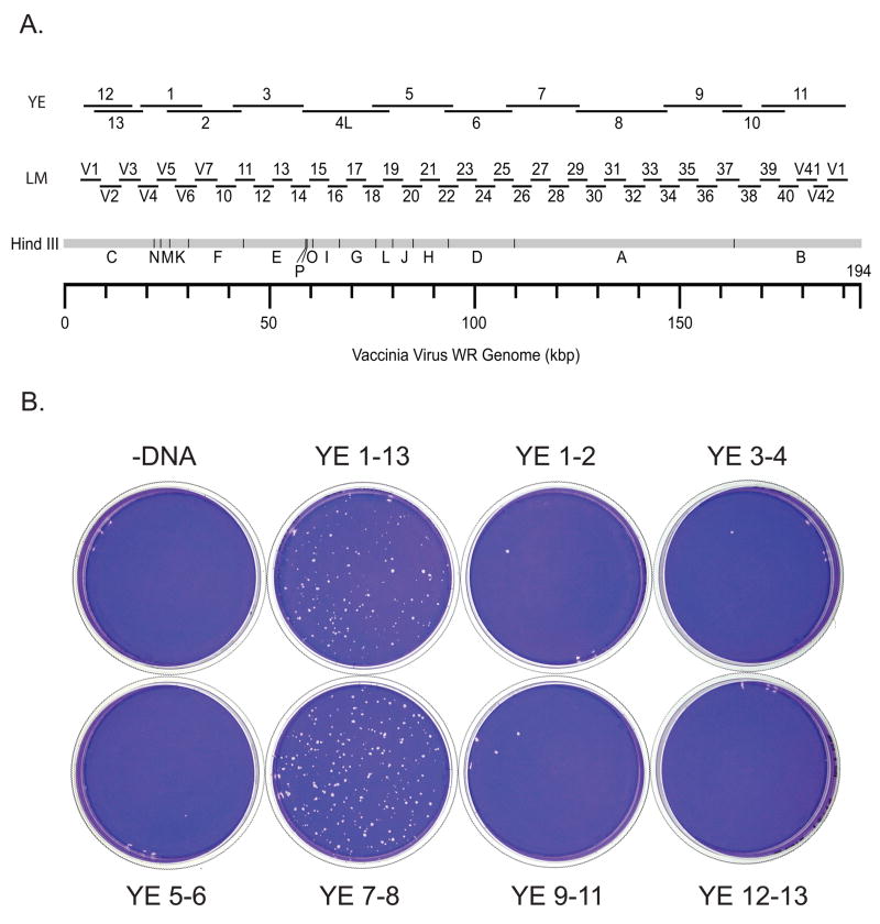
Fig. 1. Coarse marker rescue mapping of Cts13.
A) A HindIII map of the vaccinia genome showing the positions of large sized (YE) PCR fragments and intermediate sized (LM) PCR fragments. B) Marker rescue of Cts13. Dishes were infected with Cts13, transfected with pools of PCR fragments as indicated, incubated at 39.7°C for 4 days, and stained with crystal violet.

