
Fig. 1.
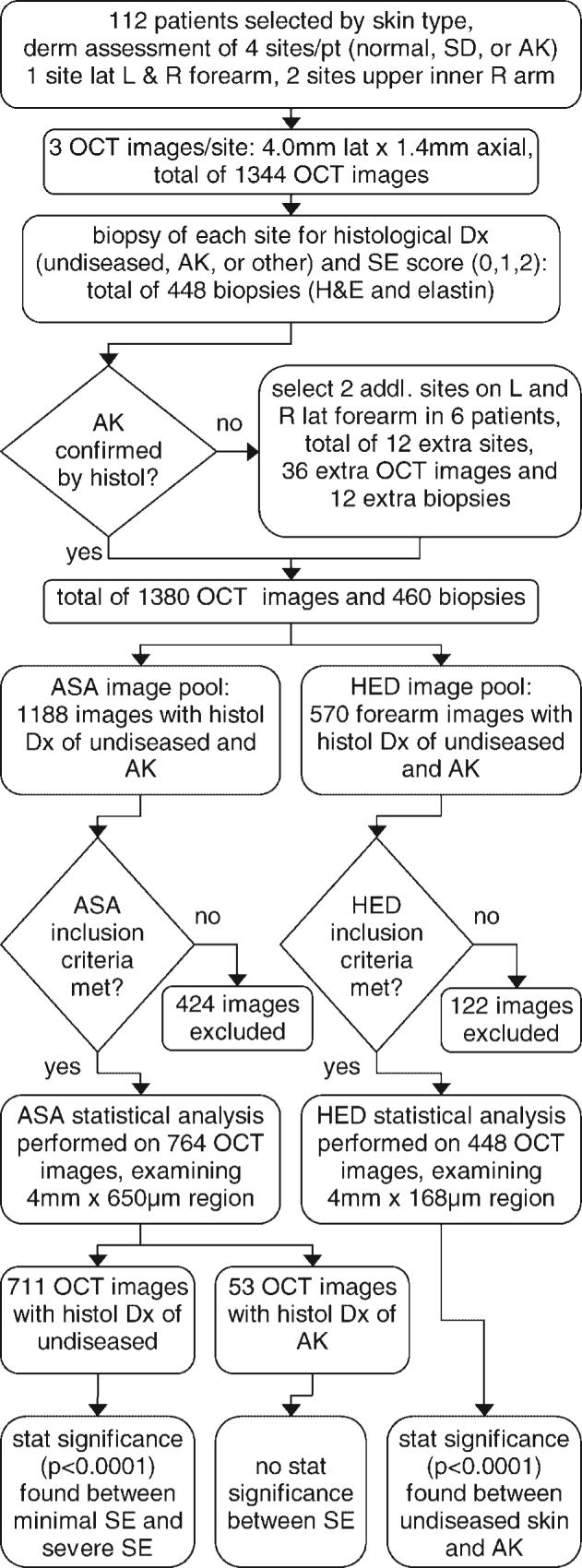
Flow chart showing the major steps in the study, including numbers of patients, images, and biopsies obtained, types of statistical image analysis, images excluded, and analysis results. Exclusion criteria for ‘average skin attenuation’ statistical analysis was (1) instrument or operator error resulting in failure to acquire images with proper settings or imaging depth, and (2) severe artifacts such as saturation or shadowing from hair/skin debris. Exclusion criteria for ‘horizontal edge detection’ was the same plus (3) severe artifacts from the surface detection procedure. addl, additional; AK, actinic keratosis; ASA, average skin attenuation; derm, dermatological; Dx, diagnosis; histol, histology; H&E, Hematoxylin & Eosin stain; HED, horizontal edge detection; L, left; lat, lateral; OCT, optical coherence tomography; pt, patient; R, right; SD, sun damaged; SE, solar elastosis; stat, statistical.