
Fig. 3.
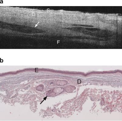
a: Example OCT image of upper inner arm of a 74-year-old male. Image shows high penetration depth, with clearly visible layers of epidermis, dermis, and subcutaneous fat, and high-contrast dermal structures. S, stratum corneum; E, epidermis; D, dermis; F, subcutaneous fat. Arrow points to hair follicle. b: Histology from same upper inner arm site as (a), with a histological diagnosis of undiseased and an SE score of 0. Arrow points to hair follicle. Symbols defined in (a).