
Fig. 4.
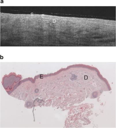
a: Example OCT image of minimal sun damaged lateral forearm of a 56-year-old female. This image shares similar epidermal characteristics the sun protected skin image (Fig. 3a), however there is lower grayscale contrast in the dermis (less visibility of dermal structures), and the subcutaneous fat layer is generally not seen. Symbols defined in Figure 3a. b: Histology from same site as (a), with a histological diagnosis of undiseased and an SE score of 0. Symbols defined in Figure 3a.