
Fig. 5.
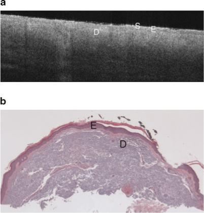
a: Example OCT image of severely sun damaged lateral forearm of a 65-year-old female. The image penetration depth is lower than seen in the minimal sun damaged skin image (Fig. 4a). Symbols defined in Figure 3a. b: Histology from same site as Figure 5a with a histological diagnosis of undiseased and an SE score of 2. Symbols defined in Figure 3a.