
Fig. 6.
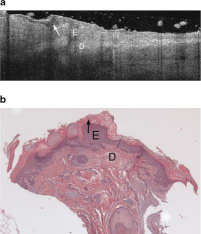
a: Example OCT image of an AK on the lateral forearm of a 77-year-old male. This AK image includes a thick dark band in the stratum corneum, which appears to be correspond to the thick keratin layer seen in corresponding histology. b: The vertical shadowing is caused by skin flaking within the keratinized region. Strong signal attenuation in the dermis is seen, similar to severely sun damaged sites (Fig. 5a). E, epidermis; D, dermis. Arrow points to keratinized region. b: Histology from same site as (a), with a very thick keratin layer, histological diagnosis of AK and an SE score of 2. Symbols defined in (a).