
Fig. 7.
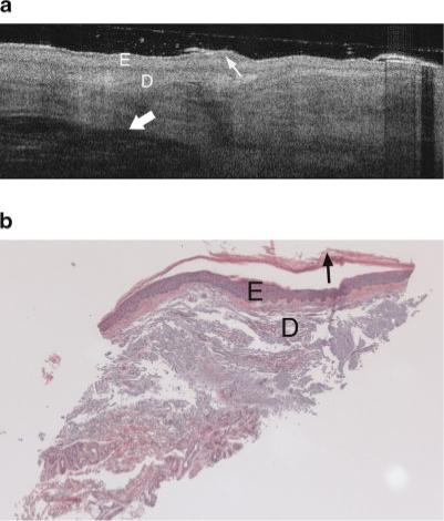
a: Example OCT image of an AK on the lateral forearm of a 74-year-old male. An increase in epidermal thickness compared to undiseased skin is seen in both OCT image and histology. Dermal structures such as a hair follicle are clearly seen (thick arrow), although wavy banding in the dermis is distinctly unusual compared to the undiseased skin case (Fig. 4a,b). Thin arrow points to keratinized region. Symbols defined in Figure 6a. b: Histology from same site as (a), with a thickened epidermis, histological diagnosis of AK and SE score of 2. Symbols defined in Figure 6a.