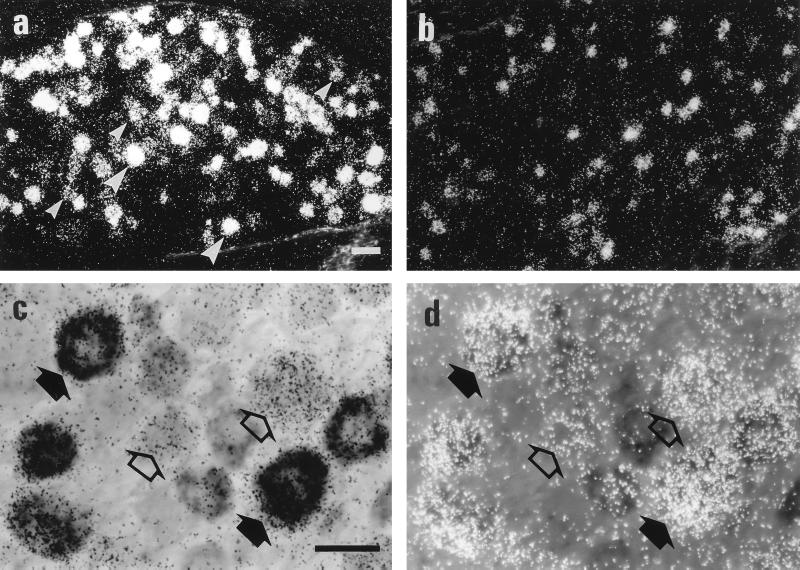
Figure 1.
Darkfield micrographs of sections of nodose ganglia hybridized for CART from normal (a) and ipsilaterally vagotomized (b) rats. Large arrowheads indicate strongly labeled neuron profile, and small arrowheads indicate weakly labeled profiles in a. (For a and b, bar in a = 50 μm.) Brightfield micrographs (c and d) show a section of rat nodose ganglion hybridized simultaneously for CART (silver grains) and CCKA receptor mRNA (dark precipitate) viewed without (c) or with (d) epiillumination. Note CART signal in all CCKA receptor mRNA-labeled neuron profiles (filled arrows), as well as several CART single-labeled profiles (open arrows). (For c and d, bar in c = 25 μm.)

