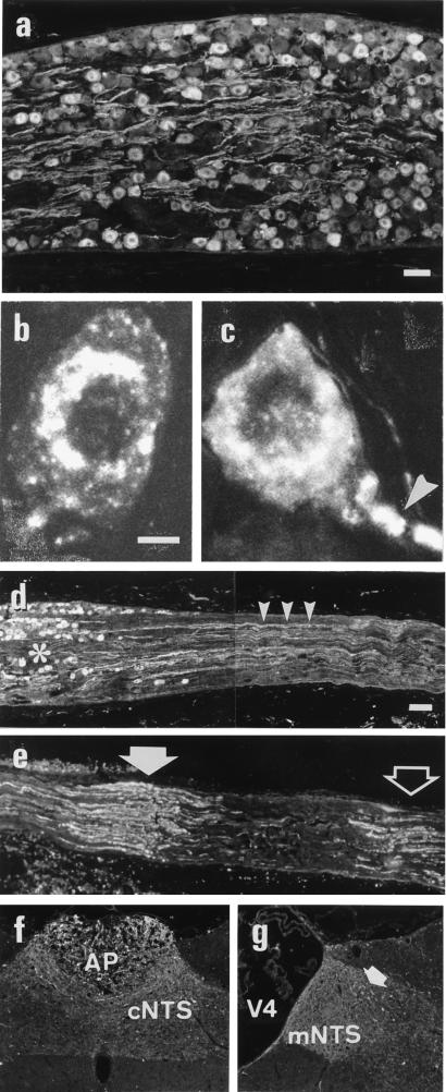
Figure 4.
Immunofluorescence (a and d–g) and confocal (b and c) micrographs of sections of rat control (a and b) and ipsilaterally vagotomized (c) nodose ganglia, normal (d) and pinched (e) rat peripheral vagus nerve, area postrema (f), and a more rostral section of the brain stem (g) stained with antiserum against CARTp. Stained fibers are seen both in the nodose ganglion (a) and vagus nerve (small arrowheads in d). Note accumulation of CARTp-LI in the cell body and the initial segment of vagotomized cell body (large arrowhead in c), as well as on the proximal (large filled arrow in e) and distal (large open arrow in e) side in the vagus nerve. Small filled arrows in g indicate CARTp-immunoreactive (-ir) cell bodies in the NTS. The asterisk in d indicates nodose ganglion. AP, area postrema; cNTS, commissural part of the NTS; medial part of the NTS; V4, fourth ventricle. (a, Bar = 50 μm for a; b, bar = 5 μm for b and c; d, bar = 100 μm for d and e, and 200 μm for f and g.)

