Figure 2.
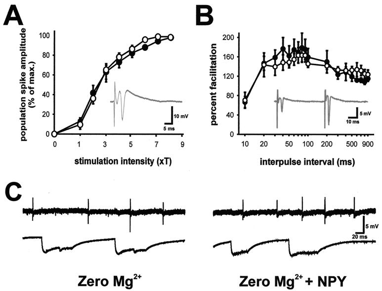
Synaptic function and epileptiform-discharge activity in hippocampal slices from Y5R−/− mice. (A) Input–output curves of population spike responses recorded in the CA1 pyramidal cell region (see Inset) to stimulation of the Schaffer collaterals. Threshold for stimulation was defined for each slice as the minimum current required to elicit a detectable population spike; the x axis shows stimulus intensity in terms of threshold multiples. Responses were normalized with respect to maximum population spike amplitude to allow averaging of responses from wild-type (○) and Y5R−/− (●) mice. (B) Plot of paired-pulse facilitation (amplitude of population spike response to second stimulus divided by amplitude of the response to first stimulus) in the CA1 pyramidal cell region (see Inset) for hippocampal slices from wild-type and Y5R−/− mice. Stimulation intensity was set at four times the threshold. (C) Spontaneous epileptiform burst-discharge activity in a hippocampal slice from a Y5R−/− mouse during perfusion with 0-Mg-ACSF (Left) and 20 min after perfusion with 0-Mg-ACSF containing 1 μM human NPY (Right). Field recordings were obtained from hippocampus (CA1 pyramidal cell layer, Upper) and neocortex (layer II–III, Lower).

