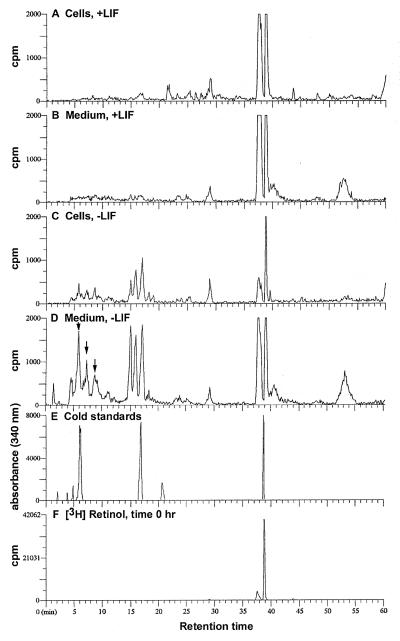
Figure 1.
Metabolism of [3H]retinol by CCE ES cells. CCE wild-type ES cells were cultured in DMEM supplemented with 10% FCS (30). Cells were cultured for 4 days plus or minus 1,000 units/ml LIF (Life Technologies). LIF was added every 24 h to the +LIF samples. After 4 days, new DMEM containing 5% FCS and 50 nM [3H]retinol was added for various times (16 h is shown). [The concentration of nonradioactive retinol in this 5% FCS was ≈50 nM (X. Guo and L.J.G., unpublished data).] The retinoids were extracted (16) and separated by HPLC (21). The identities of the retinoids were determined by coelution of the radiolabeled samples with known retinoid standards included in the samples and UV absorption spectra comparison of nonradiolabeled samples, analyzed via photodiode array, with known standards. Radiolabeled retinoids extracted from ES cells (A) or from the medium (B) of ES cells cultured with LIF for 4 days and with 50 nM [3H]retinol for 16 h. Radiolabeled retinoids extracted from cells (C) or from the medium (D) of ES cells cultured for 4 days without LIF, followed by culture with 50 nM [3H]retinol for 16 h. The all-trans-4-hydroxyretinol elutes at 15.0 min, 13-cis-4-hydroxyretinol elutes at 15.9 min, and the all-trans-4-oxoretinol elutes at 16.8 min. The arrows in D indicate unidentified, more polar [3H]retinol metabolites in the medium of cells −LIF. (E) Retinoid standards are as follows: all-trans 4-oxoRA, 6.2 min; all-trans-4-oxoretinol, 16.8 min; all-trans RA, 20.6 min; all-trans retinol, 38.7 min. (F) [3H]Retinol extracted at time zero from a control plate including medium and FCS but no cells. This experiment, including multiple time points, was performed five times with similar results.

