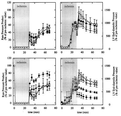
Figure 2.
Percentage changes in RPP (Left) and LVEDP (Right) in control and β-Tx hearts during 28-min ischemia and 40-min reperfusion. (Upper) Comparisons among the three genotypes used for controls (open square for −/+; filled square for +/−; open diamond for −/−). (Lower) Comparisons between pooled control (open symbols) and β-Tx (filled symbols) hearts treated with (triangles) or without (circles) CH. Data are shown as means ± SEM. The ischemia period is indicated by the shaded area.

