Figure 4.
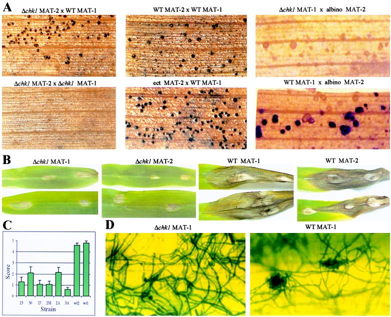
Effect of deletions at CHK1 on the ability to mate and to cause disease symptoms. (A) Mating assays. (Upper Left) chk1–23 (MAT-2) × WT (MAT-1). (Upper Center) Control WT cross. (Upper Right) chk1–2A (MAT-1) × CB12 (albino, MAT-2). (Lower Left) chk1–23 (MAT-2) × chk1–2A (MAT-1). (Lower Center) Ectopic integration strain 20 (MAT-2) × WT. (Lower Right) WT (MAT-1) × CB12. Pseudothecia are visible as dark spots or lacking pigment (albino). (B) Pathogenicity assays on corn leaves. Photographs show typical lesions formed by WT and Δchk1 strains at 8 days after inoculation. First and second photographs show Δchk1 mutants: strain 2M, an ascospore isolate of mating-type MAT-1 from a backcross of strain 30 (chk1–30 MAT-2) to WT; and strain 27 (chk1–27 MAT-2). Third and fourth photographs show WT. (C) Quantitative analysis of disease symptoms: lesions were scored on a scale of 0–5 (0 = no lesion; 1 = decolorization or necrotic lesion <1 mm; 2 = necrotic lesion 1–2 mm; 3 = 2–3 mm; 4 = 4–5 mm; 5 = severe lesion >5 mm) at 3 days after inoculation. Bars indicate SEM of a total of 15–18 replicate spots from three different plants. Differences between each mutant and wild type are significant (Student’s t test, P < 0.0002). Numbers on the x axis are strains in Fig. 2B, except wt2, wt1 (MAT-2 and MAT-1), and strain 2M, which is described in B. (D) Visualization of hyphal growth on the leaf. Corn leaves were inoculated with mycelial homogenates as in B. After 48 hr, samples were fixed, stained, and photographed at ×200. (Left) Strain 3A (chk1–3A MAT-1). (Right) WT MAT-1.

