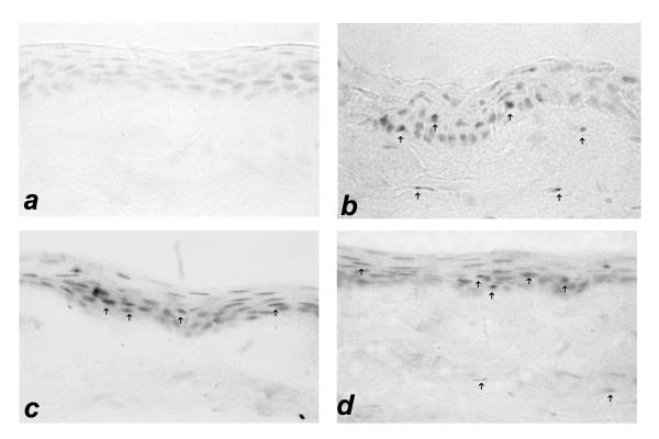
Figure 1.
Immunolocalization of MMP-1 in intact corneal epithelium at 48 hours is shown for each treatment group. Panel a: artificial tears; panel b: Ciprofloxacin 0.3%; panel c: Ofloxacin 0.3%; panel d: Levofloxacin 0.5%. A photomicrograph of the artificial tear group shows negative corneal staining for MMP-1. However, intense corneal epithelial and superficial stromal expression of MMP-1 was identified in intact corneal epithelium groups treated with fluoroquinolone eye drops (panel's b-d). The presence of MMP-1 was detected mostly in the corneal epithelial cells (arrows). (Immunohistochemistry, ×400 magnification).

