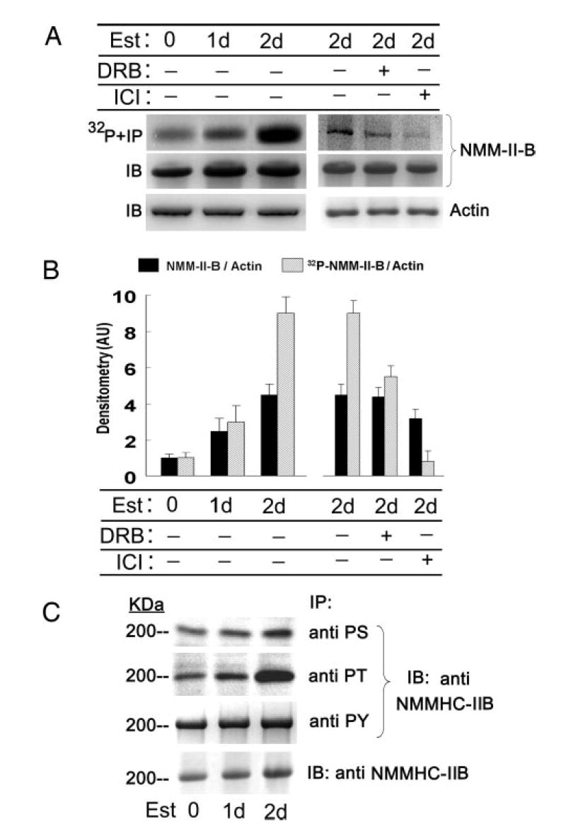
Fig. 6.

Estrogen (Est) effects on phosphorylation of NMM-II-B. CaSki cells were shifted for 3 d to steroid-free medium and treated with 10 nM 17β-estradiol for the durations indicated in the figure. In some experiments cells were also cotreated with DRB (10 μM, 6 h before assays) or ICI-182,780 (ICI, 10 μM, 1 d before assays). A, upper and middle panels, Cells were labeled with [32P]orthophosphate; lysates were fractionated on 6% PAGE and immunoprecipitated (IP) with the anti-NMMHC-II-B antibody. Lower panel, Lysates were fractionated on 10% PAGE and immunoblotted with anti-β-actin antibody. B, Densitometry of the data in A (means ± SD of three experiments). AU, Arbitrary units. C, Experiments were done as in A, except that cell lysates were immunoprecipitated with antiphosphoserine (PS), antiphosphothreonine (PT), and antiphosphotyrosine antibodies and immunoblotted (IB) with the anti-NMMHC-II-B antibody. Lower panel shows Western immunoblots (IB) of cell lysates with anti-NMM-II-HC-B antibody. The experiment was repeated twice with similar trends.
