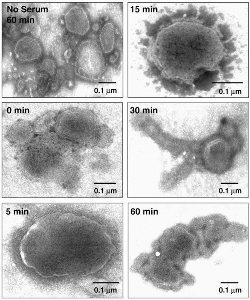
Fig. 5.

Complement induces lysis of MuV particles. Purified MuV particles were incubated for the indicated times at 37 °C with NHS and then analyzed by negative staining and EM. Note that MuV particles are not massively aggregated as seen in Fig. 4 for SV5. The solid bar in each figure represents 0.1 μm. The no serum sample represents control virions incubated at 37 °C for 60 min without serum.
