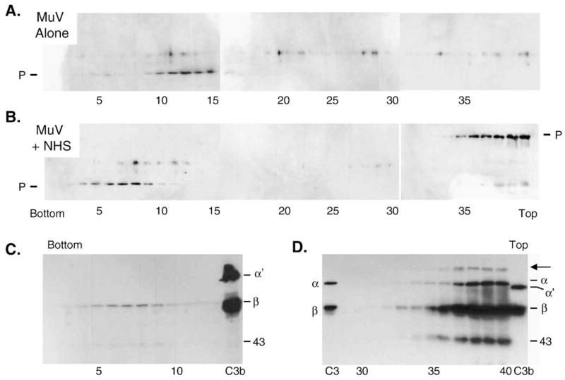
Fig. 6.

Sucrose gradient analysis of NHS-treated MuV particles. Purified MuV was incubated alone (panel A) or with NHS (panel B) for 60 min at 37 °C and then analyzed by centrifugation through 15–60% sucrose gradients. Fractions were collected and analyzed for virion proteins by western blotting with antiserum raised against the SV5 P protein which cross-reacts with MuV P protein (top and middle panels). A cross-reactive band of unknown origin is seen above the position of the P protein. C) Fractions 2–13 and 30–40 from the NHS-treated virion gradient were also analyzed by Western blotting for the presence of C3. The arrow indicates a high molecular weight protein that is not found in purified proteins in the marker lanes containing fragments of C3 and C3b.
