
Fig. 4.
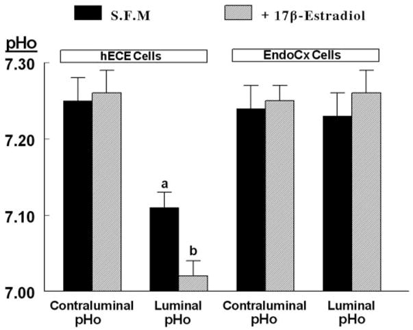
Effects of estrogen on changes in pHo. hECE cells were plated on filters and shifted to steroid-free medium for 1 d and then maintained in the same medium for an additional 2 d in the absence (S.F.M) or presence of 10 nM 17β-estradiol (added to both the luminal and contraluminal solutions) (+17β-estradiol). Determinations of pHo were described in the text. Shown are means (± SD) of three to five repeats per point of pHo determinations 30 min after mounting filters for assays. a, P < 0.01, compared with contraluminal pHo, hECE cells (S.F.M.); b, P < 0.01, compared with luminal pHo, hECE cells (S.F.M.).