
Fig. 5.
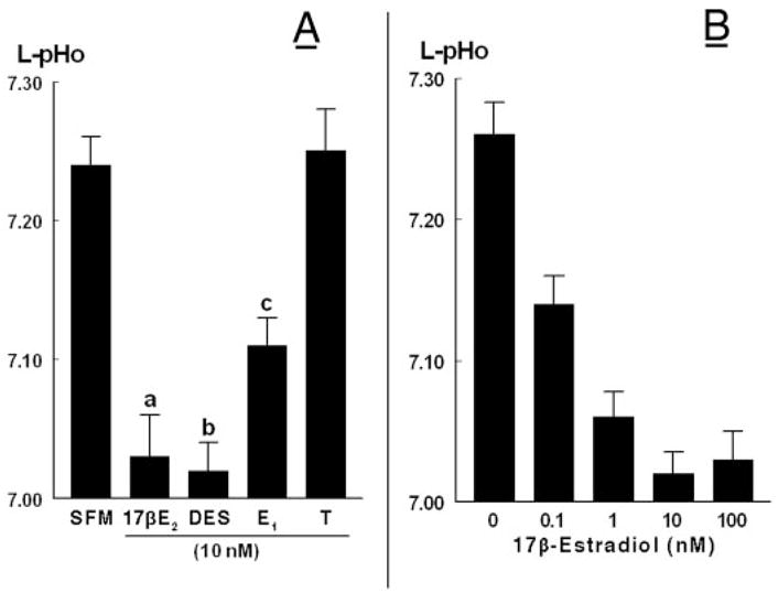
A, Specificity of estrogen-induced decrease of L-pHo. Experiments were done as in Fig. 4. hECE cells were grown in steroid-free medium (SFM) and treated with one of the following hormones (all added at 10 nM to both the luminal and contraluminal solutions): 17β-estradiol (17βE2), diethylstilbestrol (DES), estrone (E1), or testosterone. Shown are means (± SD) of L-pHo determinations in three to five repeats. a, b, c, P < 0.01, compared with SFM. B, Dose-response effect of 17β-estradiol. Shown are means (± SD) of three repeats per point. The decrease in mean (L-pHo) vs. (17β-estradiol) was significant (P < 0.01).