
Fig. 7.
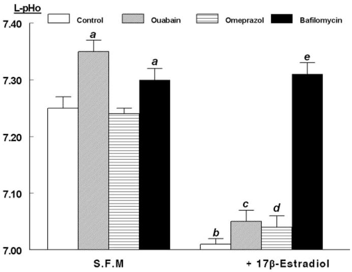
Modulation of estrogen decrease in L-pHo by ATPase inhibitors. Experiments were done as in Fig. 4. hECE cells were grown in steroid-free medium (S.F.M.) in the absence or presence of 10 nM 17β-estradiol as well as with one of the following drugs (all added 30 min before assays): ouabain (1 μM, added to the contraluminal solution), omeprazole (100 μM, added to both the luminal and contraluminal solutions), and bafilomycin A1 (1 μM, added to the luminal solution). Shown are means (± SD) of L-pHo determinations in three repeats per point 30 min after mounting filters for assays. a, P < 0.01, compared with control (C), S.F.M. group; b, c, and d, P < 0.01, compared with respective treatments in S.F.M. group; c, P < 0.02, compared with C, 17β-estradiol group, but P > 0.1, compared with omeprazole, 17β-estradiol group; e, P < 0.01, compared with b–d, and P < 0.01, compared with C, S.F.M. group.