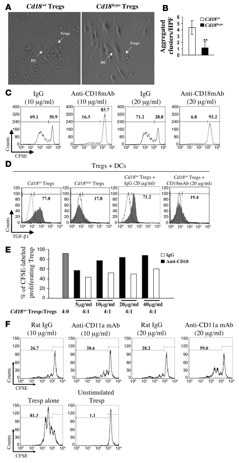
Figure 4. Reduced CD18 function disrupts cell-cell contacts between DCs and Cd18hypo Tregs from PL/J mice, which impairs specific allogeneic Treg expansion and activation.
(A) Representative pictures of cluster formation of allogeneic DCs with Tregs derived from either Cd18wt mice or Cd18hypo mice are shown. Original magnification, ×40. (B) Cluster formation between allogeneic DCs and Tregs of different genotypes from Cd18wt mice and Cd18hypo mice was assessed by counting aggregated clusters/HPF in 100 randomly selected HPFs. Cluster formation with allogeneic DCs was substantially reduced for Tregs derived from Cd18hypo mice compared with Tregs from Cd18wt control mice. **P = 0.0029, using Student’s t test. (C) Increased neutralizing mAb against CD18 resulted in decreased proliferative response of specific allogeneic Tregs in MLRs. Numbers on the top left of C and F indicate the percentage of CFSE-labeled proliferating cells. Numbers on the top right of C and F indicate the percentage of undivided CFSE-labeled cells. (D) Increased TGF-β1 expression by Cd18wt Tregs was observed in MLRs. Neutralizing mAb against CD18 in MLRs resulted in a dramatic decrease in TGF-β1 expression compared with isotype-matched control antibody. Gray region, TGF-β1 expression; white region, normal goat IgG control for TGF-β1 staining. Numbers on the top of D indicate the percentage of CFSE-labeled proliferating cells. CD4+CD25+CD127– Tregs were purified from 4 pooled spleens of Cd18wt PL/J mice and cocultured with irradiated allogeneic DCs in the presence of 500 units/ml recombinant murine IL-2 and various concentrations of anti-CD18 mAb (E), or anti-mouse CD11a mAb (F), or isotype-matched IgG for 7 days. Tregs were then separated from allogeneic DCs by CD11c MACS beads, extensively washed 3 times with PBS, and mixed at a ratio of 1:4 with Cd18wt Tresp cells. After 3 days of culture, cells were harvested and analyzed by flow cytometry. One representative experiment out of 3 or 4 independent experiments is shown.

