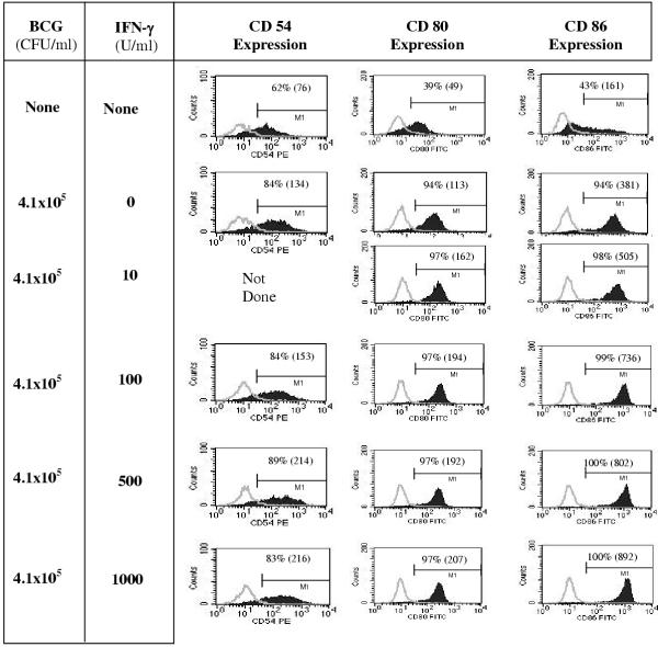
Figure 1.
IFN-γ in combination with BCG stimulates enhanced DC surface expression of CD54, CD80, and CD86 DC, derived from 6-day monocyte cultures with GM-CSF and IL-4, were left untreated, matured with 4.1 × 10⁵ CFU/ml of BCG alone, or in combination with various concentrations of IFN-γ for an additional 2 days in the presence of GM-CSF and IL-4. Cells were harvested and analyzed by flow cytometry for CD54, CD80, and CD86 expression. DC-gated (based on light scatter properties) data are shown (from a representative experiment out of three similar experiments). Isotype control staining is overlaid and shown by the light gray curve. Percent positive cells are shown in each panel and mean fluorescence intensity indicated within parentheses.

