
Figure 2.
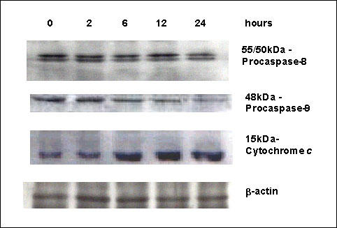
Western Blot analysis of apoptotic proteins in SPD-treated cells. Proteins from MCF-7 cells treated with 10-6 M SPD for the indicated times were resolved on 12% SDS-PAGE and submitted to Western Blotting with an anti-procaspase-8 antibody. Two bands were observed, corresponding to the uncleaved 55/50-kDa procaspase-8 isoforms. The active p18 subunit was not detected. Samples were also detected for procaspase-9 with an anti-procaspase-9 antibody (Clone B40). The anti-caspase-9 antibodies recognized the proenzyme; and the decrease of this band indicated activation of caspase-9. When proteins from cytosolic fractions of MCF-7 cells treated with 10-6 M SPD were resolved on 15% SDS-PAGE and submitted to immunoblotting with the cytochrome c antibody (Clone 7H8.2C12), increasing amounts of cytochrome c were detected in the cytosol in a time-dependent manner. All blots were then washed and reprobed with β-actin to confirm equal loading.