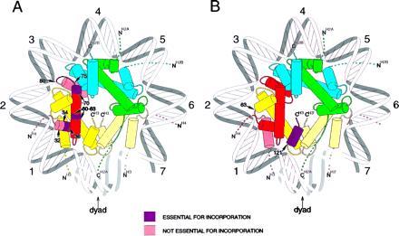
Figure 6.

Summary of role of domains of H3 and H4 in nucleosome assembly. (A) Histone H4; (B) histone H3. Histone H4 is shown in red, H3 in yellow, H2B in blue, and H2A in green. Mutated or deleted regions are as indicated. The view shown is down the superhelical axis of the DNA in the nucleosome. For simplicity, the DNA is shown as a uniform superhelix. The helical turns are numbered relative to the dyad axis (0). Only one complete heterotypic tetramer of H2A, H2B, H3, and H4 is shown. The second H3 molecule is indicated in a pale yellow to indicate the site of dimerization across the dyad axis. N and C termini of the histones are indicated; the dashed lines indicate the N-terminal tails, the exact path of which is not known at this time.
