
Figure 5.
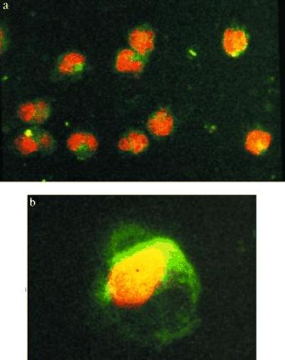
Confocal laser microscopy. After incubation during 15 min in presence of FITC–SOD (concentration, 150 units/ml), the surface of the monocytes show green fluorescence (a). After 3 h, FITC fluorescence could also be seen in the cytoplasms upon focusing through the cell. In b, a 1-μm-thick focal section taken in the middle of the cell. No labeling of the nucleus is seen.