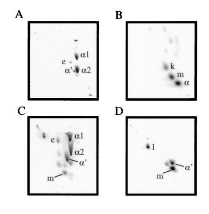
Figure 1.

2D TLC analysis of M. smegmatis (A), M. tuberculosis (B), M. smegmatis transformed with a cosmid carrying the mma locus (C), and M. smegmatis transformed with a plasmid carrying only the 6.4-kb EcoRI fragment with cma1 homology (D). The corresponding mycolate structures are shown in Fig. 2. From left to right, silica gel without silver ion impregnation (developed twice with 9:1 hexanes:ethyl acetate), from top to bottom, with silver ions (developed thrice with the same solvent). e, Epoxymycolate; k, ketomycolate (structure not shown); m, methoxymycolate; α, α mycolate from M. tuberculosis (structure not shown).
