
Figure 1.
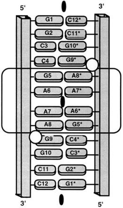
Schematic diagram of the GAAA duplex, as characterized in crystals of the dodecamer ribonucleotide r(GGCCGAAAGGCC). The phosphodiester backbone is depicted as a rectangular tube and the bases as ovals (purines, large; pyrimidines, small). The crystallographic twofold axis relating the first strand (residues 1–12) to the second strand (residues 1*–12*) is indicated as a blackened oval. Because of this exact symmetry only 6 base pairs are unique, i.e., base pairs 1–6·7*–12* are the same as pairs 1*–6*·7–12. The 4 terminal base pairs on either end are formed by standard Watson–Crick pairing, while the middle 4 bases form an internal loop of nonstandard pairs as indicated by the boxing. Bound Mn2+ ions are represented by circles.