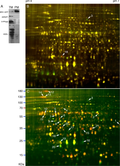
Fig. 6.
2-D DIGE analyses of BR-regulated plasma membrane proteins. A, immunoblots of microsomal (TM) and plasma membrane (PM) proteins of the det2 mutant expressing BRI1-GFP fusion protein. Three micrograms of microsomal or plasma membrane proteins were separated by SDS-PAGE. Western blots were probed using antibodies against different membrane marker proteins (BRI1-GFP, PM; AtRGP1, Golgi; vacuolar H+-translocating pyrophosphatase (V-PPase), tonoplast; HDEL, ER). B and C, 2-D DIGE image of plasma membrane proteins from 7-day-old det2 seedlings treated with BL for 2 (B) or 24 h (C). BL-treated PM proteins were labeled by Cy5, and mock-treated PM proteins were labeled by Cy3. Spots identified by LC-MS/MS are marked by arrows and numbers (listed in Table II).

