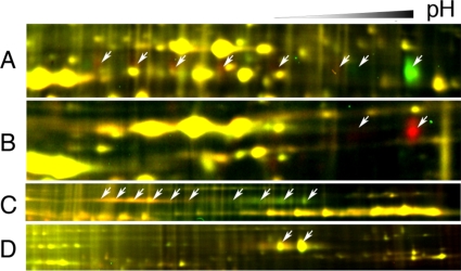
Fig. 7.
BAK1 is posttranslationally modified after BR treatment. A, zoomed-in view of the region of Fig. 6B containing BAK1 (spot 3). Arrows show potential shifted BAK1 spots that disappear (green spots) or appear (red spots) after BL treatment. B, BAK1 spots disappear in bak1-1 mutant. PM proteins were isolated from wild type and T-DNA knock-out mutant bak1-1 and labeled with Cy5 and Cy3, respectively. Arrows show the BAK1 spots. C and D, plasma membrane proteins isolated from transgenic plant expressing the BAK1-GFP fusion protein in Columbia (C) or bri1-5 (D) were labeled with Cy5 (BL-treated) or Cy3 (mock-treated) and analyzed by 2-D DIGE. Arrows show the BAK1-GFP protein spots.

