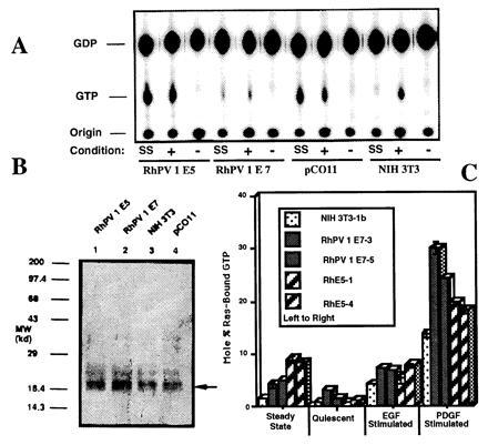
Figure 1.

Determination of p21ras-bound guanine nucleotides from immunoprecipitates of normal and transformed NIH 3T3 cells. (A) Labeled cells were disrupted following growth under steady-state (SS), EGF-stimulated (+), or quiescent (−) conditions. Anti-p21ras antibody was used to immunoprecipitate p21ras and bound nucleotide. Following dissociation, nucleotides were separated using polyethyleneimine-cellulose chromatography and autoradiographed. The positions of the origin, GDP, and GTP are shown. (B) Anti-p21ras antibody Western blot of anti-p21ras immunoprecipitated cell lysates. Arrow indicates position of p21ras. (C) Independent additional clones of NIH 3T3 (clone 1b), RhPV 1 E7 (clones 3 and 5), and RhPV 1 E5 (clones 1 and 4) cells were tested in duplicate assays for Ras activation under various conditions as indicated with standard error bars shown.
