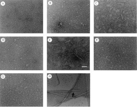
Figure 2.

In vitro assembly of pilus-like or tip fibrillum-like fibers after chaperone uncapping. Panels A–H are electron micrographs of chaperone–subunit complexes following rapid freeze-thaw conditions. PapD–PapA complexes (a mixture of DA, 1:1 PapD–PapA, and DA2, 1:2 PapD–PapA) before (A) and after three (B) or 10 (C) freeze-thaw cycles. (D and E) The PapD–PapA complexes after 10 freeze-thaw cycles in the presence (D) or absence (E) of a 10-fold excess of free PapD. Freeze-thaw treatment of purified PapD-PapE complex and PapD-PapK complex is shown in F and G, respectively. H is an electron micrograph of purified P pili showing the composite architecture of the fiber: a tip fibrillum (→) joined end to end to a pilus rod. Magnification bar in E = 500 Å.
