
Figure 6.
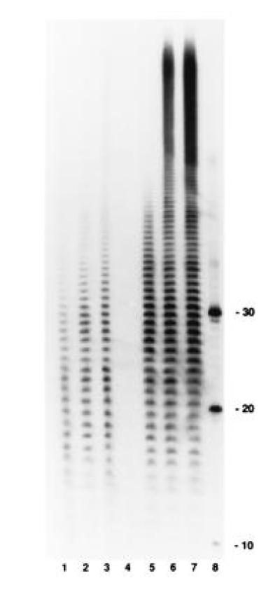
Poly(A) tails formed during in vitro mRNA decay. Polysomes were incubated in the presence of [α-32P]ATP. Samples were removed at various times, deproteinated, and then digested exhaustively with RNase A and RNase T1 as described. An autoradiogram of a 12% polyacrylamide gel is shown. Lanes 1–3, SK5667 (PAP I+) at 2, 5, and 10 min, respectively; lane 4, SK8964 (PAP I−) at 10 min; lanes 5–7, SK8964 (PAP I−) with 0.4 units of exogenous PAP I per reaction at 2, 5, and 10 min, respectively; and lane 8, 10-, 20-, and 30-mers of oligo(dA) end-labeled with [γ-32P]ATP.