
Figure 5.
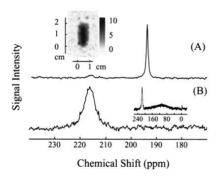
129Xe NMR spectra in fresh human blood obtained using laser-polarized 129Xe in A, 20% Intralipid solution and B, Fluosol. The 129Xe NMR signal at a chemical shift of 216 ppm is characteristic of xenon in the RBC, and the signal at 194 ppm is of xenon in the plasma/lipid mixture (A). (A Inset) A two-dimensional projection MRI image of 129Xe dissolved in the mixture of lipid and fresh human blood. The 128 pixel × 64 pixel image was obtained by the Echo Planar Imaging (26, 27) method on a Quest 4300 (Nalorac Cryogenics, Martinez, CA) spectrometer. The diameter of the sample tube was 1 cm, and the solution occupied a region of 2 cm in height. (B Inset) The broad peak centered around 110 ppm is due to xenon in Fluosol. NMR experiments, except the imaging experiment as mentioned above, were performed on a Bruker AM-400 spectrometer.