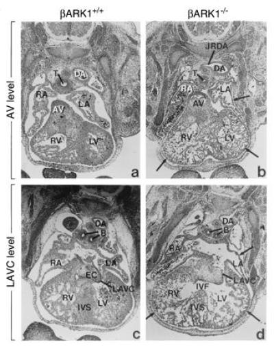
Figure 4.

Cardiac abnormalities in βARK1 homozygote embryos visualized with hematoxylin- and eosin-stained sections of whole embryos. Two sections each are shown at the aortic valve and left atrioventricular canal level. Structures are labeled as follows: B, bronchi; DA, descending aorta; EC, endocardial cushion; IVF, interventricular foramen; IVS, interventricular septum; JRDA, junction of the right dorsal aorta; LA, left atrium; LAVC, left atrioventricular canal; LV, left ventricle; RA, right atrium; RV, right ventricle; T, trachea.
