Figure 5.

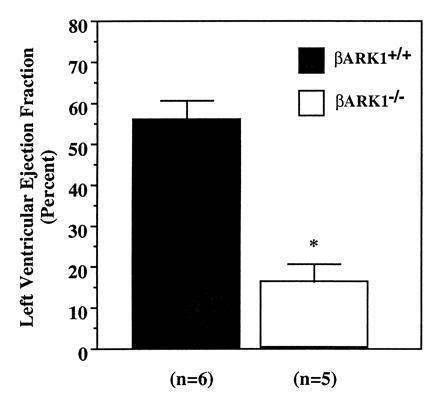
In vivo assessment of cardiac ejection fraction in E12.5–E13.5 embryos. Images were obtained on embryos with an intravital microscope and recorded at 30 frames/sec. n = the number of embryos from three different litters.

In vivo assessment of cardiac ejection fraction in E12.5–E13.5 embryos. Images were obtained on embryos with an intravital microscope and recorded at 30 frames/sec. n = the number of embryos from three different litters.