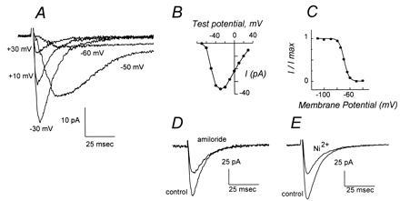
Figure 1.

Ca2+ currents of mouse spermatogenic cells are mediated by low-voltage-activated T channels. (A) Montage of whole-cell Ca2+ current traces after depolarizations of a round spermatid to the indicated membrane potentials from a holding potential of −90 mV. A slowly inactivating component of ICa after depolarization to positive potentials, diagnostic of L currents, is not apparent. (B) Current–voltage relationship for peak current amplitude elicited by depolarizations from a holding potential of −90 mV. (C) Voltage dependence of steady-state inactivation of round spermatid Ca2+ current. (D) Inhibition of peak Ca2+ current amplitude by amiloride. Traces show current elicited by depolarizations from −80 mV to −20 mV in a cell prior to (control) and after addition of 200 μM amiloride. (E) Inhibition of peak Ca2+ current amplitude by Ni2+. Traces show current elicited by depolarizations from -80 mV to -20 mV in a cell prior to (control) and following addition of 50 μM Ni2+. Current scales are indicated and differ between panels A, D, and E.
