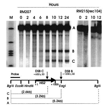
Figure 2.

Southern analysis of DSBs during meiosis at the HIS2 locus. Cells were removed from sporulation medium at the times indicated, and DNA was made, digested with BglII, and analyzed as described. The numbers above the lanes refer to the time in meiosis. The parental band A and the DSB bands B and C are illustrated in the figure. The probe was the BglII–EcoRI fragment shown on the figure. The diploid used for the wild-type lanes was RM207 (his2-xho/HIS2 rad50S/rad50S, congenic to RM169). On the right side of the figure are four time points from a congenic rec104-Δ1/rec104-Δ1 rad50S/rad50S diploid (RM215). The lane labeled M contains markers of sizes 5.2 kb, 3.9 kb, 3.2 kb, 3.0 kb, 2.9 kb, and 1.9 kb, from top to bottom. All lanes shown were run on the same gel.
