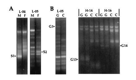
Figure 1.

Examples of differential display comparing male versus female (A, M versus F) and gut versus carcass (B, G versus C) mRNA. Amplification products were resolved on 1.4% ethidium-stained agarose gels. PCR amplification used decamer primers L-04 (5′-GACTGCACAC-3′), L-05 (5′-ACGCAGGCAC-3′), H-14 (5′-ACCAGGTTGG-3′), and H-16 (5′-TCTCAGCTGG-3′), in combination with an oligo(dT) primer. H-14 and H-16 reactions were performed in duplicate. The differentially amplified PCR fragments S1, S2, G3, G13, and G14 were isolated for further analysis.
