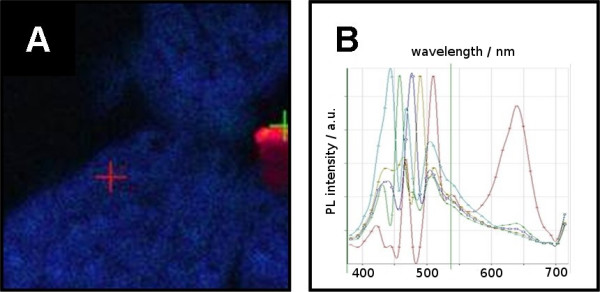
Figure 3.

A) LSM-picture of selectively labeled Arabidopsis tissue. The tissue was hybridized with five different QD-oligonucleotides of distinguishable emission wavelength. The emission of QD-labels was shifted to the strong blue. This shift appears only after hybridization. Unreacted, agglomerated QD-oligonucleotides show their initial emission behavior (red area on the right border of the picture). B) Spectral analysis of picture A. The wavelength of all hybridized QD-oligonucleotides was shifted to 460 nm. The emission of some non hybridized QD-oligonucleotides could be found at 598 nm.
