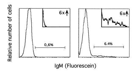
Figure 1.

Example of the analysis of surface IgM expression after IL-7 withdrawal. Clone AP-5 was cultured on the stromal layer for 3 days in the presence (Left) or in the absence (Right) of IL-7. Fluorescence-activated cell sorter analysis of IgM expression was performed as described. (Insets) An enlargement of the areas containing IgM-positive cells.
