
Figure 2.
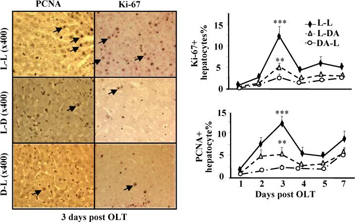
Hepatocyte proliferation is lower in the allogeneic grafts vs. the syngeneic grafts. The liver tissues from Fig. 1 were stained with anti-PCNA or anti-Ki67 antibodies. Representative photomicrographs are shown in left panel. The numbers of Ki67+ and PCNA+ hepatocytes were counted and shown in right panel. Values are shown as means ± SEM (n=3−6 as described in Fig. 1). **P<0.01, ***P<0.001 compared with values from corresponding L-L syngeneic grafts.