
Fig. 1. Deletion of GNMT leads to steatosis, fibrosis, and HCC.
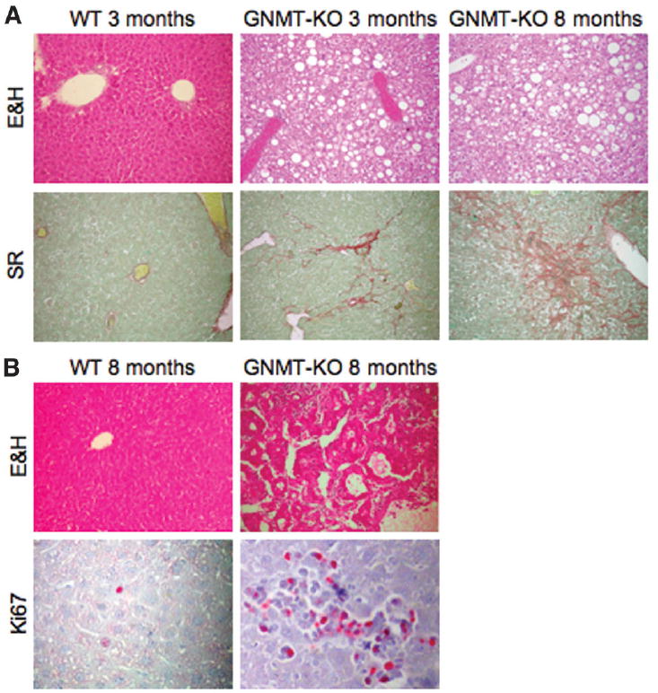
(A) At 3 months of age, macro and microvesicular steatosis (white droplets) could be seen through the hepatic lobule in GNMT-knockout (GNMT-KO) mice compared with wild-type (WT) animals. Collagen deposits (stained red) indicate moderate liver fibrosis. By 8 months of age, liver steatosis and fibrosis in mutant mice were more prominent. At least 10 animals per group were examined. (B) At 8 months of age, livers from GNMT-KO mice also had multifocal HCC. Liver cords up to 5 cells thick, lined by endothelial cells, could be seen, with occasional pseudogland formation. Staining with the nuclear proliferation marker Ki67 (red spots) indicates a higher proportion of cells in the cell cycle compared with WT cells. At least 10 animals per group were examined (E&H, eosin and hematoxylin staining; SR, Sirius red staining). Original magnification ×40.