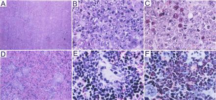
Figure 6.

Pathologic analysis of the spleens of mice injected with BCR/ABL-infected p53−/− (A–C) or p53+/+ (D–F) marrow cells. (A) Spleen of mouse injected with BCR/ABL-infected p53−/− cells (H&E stain; ×25). (B) High-power image of splenic leukemic infiltrate (H&E stain; ×350). (C) Chloroacetate esterase stain (red) to detect myeloid differentiation (Leder staining; ×350). (D) Spleen of mouse injected with BCR/ABL-infected p53+/+ marrow cells. (E) High power image of splenic leukemic infiltrate (H&E stain; ×350). (F) As in C (Leder staining; ×350).
