
Fig. 5.
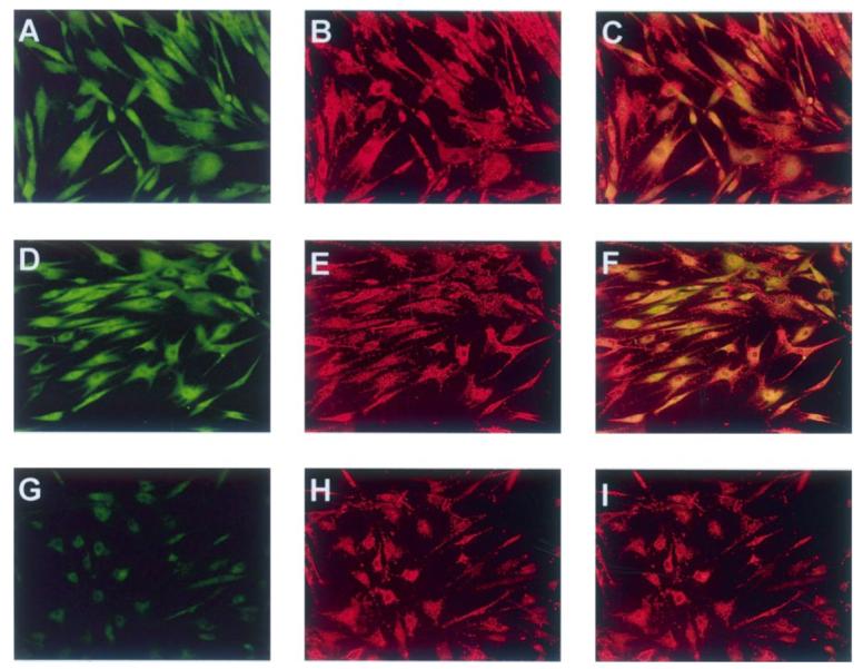
Tyrosine nitration occurs within AIDS-KS mitochondria. Cells were cultured on chamber slides, actively respiring mitochondria localized by addition of MitoTracker Red CMXRos, and the presence of 3-nitrotyrosine confirmed by antinitrotyrosine antibody. The same fields were photographed under Alexa 488 (green, 3-nitrotyrosine) and Alexa 579 (red, MitoTracker CMXRos) excitation wavelengths. Regardless of culture conditions, 3-nitrotyrosine positive staining was observed in the majority of AIDS-KS cells (A, log growth, D, 24 h sera deprivation) and mitochondria were visible as discrete, punctuate intense red staining (B, log growth, E and H, 24 h sera deprivation). Co-localization results reveal foci of yellow-orange staining consistent with 3-nitrotyrosine staining within the mitochondria (C, log growth, F, 24 h sera deprivation, I, 24 h sera deprivation co-localized with isotypic control). (All photomicrographs are at 200× image scale.)