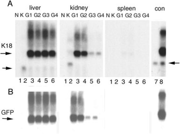
FIG. 2.

Northern blot analysis of K18iresEGFP transgenic tissues. Ten μg of total RNA was hybridized with radioactive probes for EGFP (B) and human K18 (A) sequentially. Samples for liver RNA were separated in a different gel than those for kidney and spleen. Samples from four founder lines are indicated by G1, G2, G3, and G4. N, nontransgenic mouse RNA; K, RNA from K18TG1, a low copy number human K18 transgenic mouse (Abe and Oshima, 1990); con, RNA from HR9 cells (lane 7) and the same cells transfected with the K18iresEGFP vector (lane 8). The position of the K18iresEGFP chimeric mRNA is indicated by the upper horizontal arrow. The lower arrow indicates the K18 mRNA from K18TG1. In lanes 7 and 8, the lower arrow indicates the cross-hybridization of the K18 probe with mouse K18. Overlaying the films of A and B revealed that the EGFP and major K18 signals were coincident.
