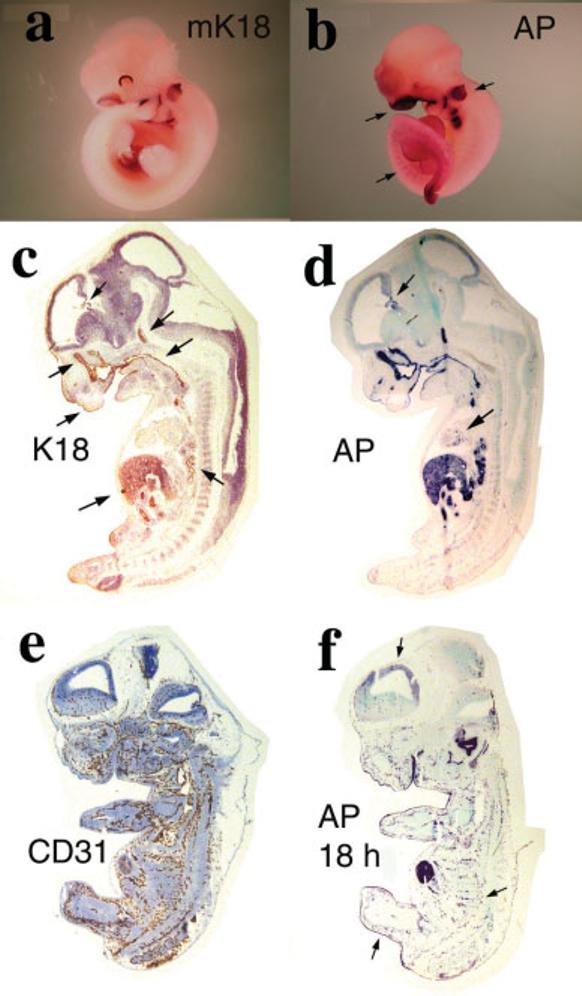
FIG. 3.

Human K18 and AP expression in K18iresAP embryos. a: ISH of E10.5 wild-type mouse embryo with mK18 riboprobe. b: AP staining of E10.5 K18iresAP embryo. Arrows point to strong staining in the otic vesicle and nasal area. Lower arrow points to weak, striated staining of the posterior trunk. c–f: Sections of E12.5 K18iresAP embryos. c: Human K18 antibody stain. Arrows point to staining of periderm, nasal epithelium, choroid plexus, pituitary, pharynx/esophagus, liver, and lung. d: Four-hour AP staining of adjacent section of c. Arrows point to staining in choroid plexus and heart. e: CD31 antibody localization of developing vasculature. f: 18-h AP staining of adjacent section to e. Arrows point to periderm, striated vascular staining, and weak neopallial cortex staining.
